

Carbon Monoxide Poisoning: Causes, Symptoms & Treatment
Medically Reviewed by Dr. Abdul Latif Saad
Known as the “silent killer,” carbon monoxide is a toxic, odorless, and colorless gas. Immediate recognition, rapid evacuation to fresh air, and prompt high-flow oxygen therapy are essential to prevent severe brain and cardiac damage.
Carbon monoxide (CO) poisoning is one of the most common and potentially lethal types of toxic gas exposures worldwide. Because carbon monoxide is completely colorless, odorless, tasteless, and non-irritating, victims are often entirely unaware of its presence until significant physiological impairment occurs — earning it the reputation of a “silent killer” [1][2].
Exposure typically occurs in enclosed or poorly ventilated spaces where fuel-burning appliances, vehicle engines, or fires generate incomplete combustion products. When inhaled, carbon monoxide binds aggressively to hemoglobin in red blood cells, severely impeding the body’s ability to transport and release oxygen to vital organs such as the brain and heart [1][5]. Early diagnosis, prompt administration of high-flow oxygen, and long-term neurocognitive follow-up form the cornerstone of effective management [2][3].
✔ Quick Summary
- ✓ Mechanism: Carbon monoxide binds to hemoglobin with an affinity 200 times greater than oxygen, forming carboxyhemoglobin (COHb) and causing severe cellular tissue hypoxia [1][5].
- ✓ Nonspecific Presentation: Early symptoms mimic viral flu (headache, dizziness, nausea, fatigue) without fever, making high clinical suspicion critical [2][6].
- ✓ Diagnostic Pitfall: Standard pulse oximeters (SpO2) cannot distinguish carboxyhemoglobin from oxyhemoglobin, yielding falsely normal readings. Diagnosis requires blood co-oximetry [1][2].
- ✓ Treatment Protocol: Immediate removal from exposure and 100% normobaric oxygen via non-rebreather mask is universal first-line therapy. Hyperbaric oxygen therapy (HBOT) is indicated for severe cases [2][3].
1. What Is Carbon Monoxide Poisoning?
Carbon monoxide poisoning is a life-threatening systemic toxicological condition resulting from the inhalation of carbon monoxide gas. When inhaled, carbon monoxide rapidly diffuses across the alveolar-capillary membrane into the bloodstream, where it competitively binds to the iron centers of hemoglobin, myoglobin, and mitochondrial cytochrome enzymes [1][5].
Because carbon monoxide’s affinity for hemoglobin is approximately 200 to 250 times greater than that of oxygen, even low ambient concentrations of the gas can displace oxygen and significantly reduce the oxygen-carrying capacity of blood [1]. Furthermore, carbon monoxide toxicity extends beyond simple hypoxemia: it triggers cellular metabolic disruption, oxidative stress, neuroinflammation, and direct myocardial injury [1][2].
Carbon monoxide binding to one of the four oxygen-binding sites on hemoglobin alters the protein structure, increasing oxygen affinity at the remaining three sites (the Haldane effect). This shifts the oxygen-hemoglobin dissociation curve to the left, preventing oxygen release to oxygen-starved peripheral tissues [1][5].
2. How Does Carbon Monoxide Poisoning Develop?
The biological development of carbon monoxide toxicity involves a multi-step cascade affecting cellular respiration and microvascular integrity:
- Carboxyhemoglobin (COHb) Formation: Inhaled carbon monoxide binds rapidly with hemoglobin to form carboxyhemoglobin, impairing systemic oxygen delivery throughout the arterial network [1].
- Myoglobin Binding & Cardiac Depression: Carbon monoxide binds to cardiac myoglobin with high affinity, impairing oxygen storage in muscle tissue. This causes acute myocardial depression, reduced cardiac output, and systemic hypotension [1][2].
- Inhibition of Mitochondrial Cellular Respiration: CO binds to cytochrome c oxidase (complex IV of the electron transport chain), halting mitochondrial ATP synthesis and forcing cells into anaerobic metabolism, lactic acidosis, and energy failure [1][5].
- Endothelial Damage & Delayed Neurological Sequelae (DNS): CO promotes neutrophil adhesion to brain microvasculature, leading to xanthine oxidase activation, lipid peroxidation, and neuroinflammatory cascades. This immunologic cascade explains why some patients develop delayed neurological sequelae weeks after initial recovery [1][3].
3. Causes of Carbon Monoxide Poisoning
Carbon monoxide is produced by the incomplete combustion of carbon-containing fuels (natural gas, gasoline, propane, wood, coal, and kerosene). Under normal operating conditions with adequate oxygen and ventilation, fuel burns to produce carbon dioxide (CO2). However, in oxygen-depleted or enclosed environments, dangerous levels of carbon monoxide accumulate rapidly [1][4].
4. Risk Factors
⚠️ Who Is Most at Risk?
While anyone exposed to carbon monoxide can suffer fatal toxicity, certain physiological groups face substantially higher clinical vulnerability:
Unborn Fetuses & Pregnant Women: Fetal hemoglobin (HbF) binds carbon monoxide with an even higher affinity than adult hemoglobin. Carboxyhemoglobin clears much more slowly from fetal blood, placing the fetus at severe risk of tissue hypoxia, brain injury, or fetal death [1][2].
Infants & Young Children: Children have higher minute ventilation rates and baseline metabolic oxygen requirements relative to body mass, causing them to absorb environmental carbon monoxide more rapidly than adults [2][4].
Older Adults & Patients with Cardiac Disease: Pre-existing coronary artery disease or cerebrovascular disease significantly reduces physiological tolerance to hypoxia. Low-level CO exposure can precipitate acute coronary syndrome, arrhythmias, or ischemic stroke [1][2].
Storm Victims & Generator Users: Power outages during severe weather frequently prompt improper indoor use of portable gasoline generators or charcoal heaters, representing a major spike in acute poisonings [4].
Firefighters & Industrial Workers: Individuals involved in firefighting or industrial processes utilizing methylene chloride (a solvent metabolized by the liver into carbon monoxide) face elevated risk [1][4].
5. Signs & Symptoms
The clinical presentation of carbon monoxide poisoning ranges from mild constitutional “flu-like” complaints to severe neurological and cardiovascular collapse. Recognizing the spectrum of symptoms is critical [2][6]:
- Tension-Type Headache: The single most common presenting symptom (occurring in over 90% of cases), typically described as a dull, continuous frontal headache [6].
- Dizziness & Lightheadedness: Caused by reduced cerebral oxygen delivery and mild intravascular hypotension [2][6].
- Nausea & Vomiting: Frequently misdiagnosed as acute viral gastroenteritis or food poisoning [4][6].
- Confusion & Ataxia: Disorientation, memory impairment, gait instability, and motor incoordination reflect central nervous system toxicity [2].
- Chest Pain & Tachycardia: Acute myocardial ischemia or palpitations caused by oxygen deprivation and myoglobin binding in heart muscle [1][2].
- Syncope & Loss of Consciousness: A high-risk indicator of severe toxicity carrying an elevated risk for long-term brain injury [2][3].
- Seizures & Coma: Severe toxic-anoxic encephalopathy requiring immediate airway protection, intubation, and intensive care [1][2].
- Delayed Neurological Sequelae (DNS): Cognitive decline, memory failure, movement disorders (Parkinsonism), and personality changes that emerge 2 to 40 days after initial recovery [1][3].
6. How Is Carbon Monoxide Poisoning Diagnosed?
Diagnosing carbon monoxide poisoning requires a high index of clinical suspicion coupled with objective blood gas co-oximetry [1][2].
- Blood Co-Oximetry (Gold Standard): Diagnosis is confirmed by measuring carboxyhemoglobin (COHb) levels in arterial or venous blood via a specialized co-oximeter. Normal non-smoker COHb levels are below 2–3%, while smokers may have baseline levels between 5–10%. A COHb level greater than 3–5% in non-smokers (or >10% in smokers) confirms exposure [1][2].
- Electrocardiogram (ECG) & Cardiac Biomarkers: 12-lead ECG and serum Troponin I/T levels must be obtained in all moderate-to-severe cases to rule out acute myocardial infarction or ischemic arrhythmias [1][2].
- Neuroimaging (CT / MRI): Brain CT or MRI may reveal bilateral globus pallidus lesions or diffuse cerebral edema in severe acute cases or patients presenting with delayed neurological symptoms [1][3].
Standard bedside pulse oximeters (SpO2) pass light at two wavelengths (660nm and 940nm) and cannot distinguish oxyhemoglobin from carboxyhemoglobin. A patient with fatal 50% carboxyhemoglobin toxicity will register a falsely reassuring SpO2 reading of 98–100%. Never rely on standard pulse oximetry to rule out carbon monoxide poisoning [1][2].
7. Differential Diagnosis
🔎 Conditions That Can Look Similar
Because carbon monoxide symptoms are non-specific, it is frequently misdiagnosed as other common medical illnesses [2][4]:
8. Treatment Options
Management of carbon monoxide poisoning centers on rapid removal from exposure, competitive displacement of CO from hemoglobin with high-concentration oxygen, and supportive care [1][2][3].
🏥 A. First-Line Treatment: 100% Normobaric Oxygen
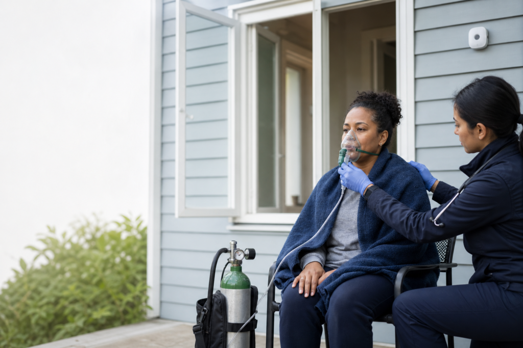
The immediate intervention for all suspected CO poisonings is 100% normobaric oxygen delivered via a tight-fitting non-rebreather mask [1][2]. On room air (21% O2), the elimination half-life of carboxyhemoglobin is approximately 320 minutes (5.3 hours). Breathing 100% normobaric oxygen reduces the COHb half-life to approximately 74 minutes, rapidly restoring systemic oxygen delivery [1][2].
💊 B. Hyperbaric Oxygen Therapy (HBOT)
Hyperbaric Oxygen Therapy involves administering 100% oxygen at 2.5 to 3.0 atmospheres of absolute pressure (ATA) inside a specialized pressure chamber. HBOT further reduces the COHb half-life to approximately 20 minutes, dissolves oxygen directly in blood plasma to nourish tissues independent of hemoglobin, and reduces lipid peroxidation and neuroinflammation [1][3].
- Key HBOT Indications: Syncope or loss of consciousness, COHb > 25%, pregnancy with COHb > 15%, evidence of acute myocardial ischemia, severe neurological deficits, or persistent confusion despite normobaric oxygen [2][3].
🏠 C. Long-Term Monitoring & Environmental Safety
Patients recovering from moderate-to-severe poisoning should undergo neurocognitive evaluation 1 to 2 months post-discharge to screen for Delayed Neurological Sequelae (DNS) [1][3]. Under no circumstances should patients return to a home or workplace until local fire authorities or gas utilities inspect and clear the property.
📚 9. Evidence in Context
The evidence base for carbon monoxide management is supported by major clinical reviews in the New England Journal of Medicine and American Journal of Respiratory and Critical Care Medicine [1][2]. While 100% normobaric oxygen is universally accepted, the precise role and optimal regimen of Hyperbaric Oxygen Therapy (HBOT) remain a subject of active discussion. A Cochrane systematic review noted trial heterogeneity regarding HBOT’s reduction of long-term Delayed Neurological Sequelae (DNS) [3]. Nonetheless, major hyperbaric medicine guidelines recommend HBOT for high-risk patients (syncope, pregnancy, cardiac ischemia, severe acidosis) when administered within 6 hours of exposure [2][3].
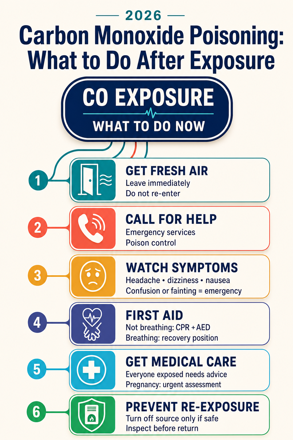
10. When to Seek Emergency Medical Help
• You suspect carbon monoxide exposure in your home or workplace
• Multiple family members or co-workers become suddenly unwell with headaches/nausea
• Anyone experiences confusion, dizziness, chest pain, or difficulty breathing
• Anyone collapses, suffers a seizure, or loses consciousness
• Pets in the building are lethargic, weak, or unresponsive
11. Frequently Asked Questions (FAQ)
Q Why don’t standard pulse oximeters work for carbon monoxide poisoning?
Standard finger pulse oximeters measure light absorption at wavelengths that cannot distinguish carboxyhemoglobin (CO bound to hemoglobin) from oxyhemoglobin (oxygen bound to hemoglobin). Consequently, a dangerously poisoned patient can show a falsely normal SpO2 reading of 99–100%. Blood co-oximetry is required [1][2].
Q What are Delayed Neurological Sequelae (DNS)?
Delayed Neurological Sequelae refer to brain impairments (memory loss, cognitive decline, personality changes, motor dysfunction) that develop 2 to 40 days after apparent recovery from acute carbon monoxide poisoning. They result from delayed neuroinflammatory pathways and brain lipid peroxidation [1][3].
Q How can carbon monoxide poisoning be prevented at home?
Install UL-listed carbon monoxide detectors on every level of your home and outside sleeping areas. Never operate gasoline generators, charcoal grills, or unvented fuel heaters inside enclosed spaces or near open windows, and have fuel-burning appliances inspected annually by qualified technicians [2][4].
References
- Rose JJ, Wang L, Xu Q, McTiernan CF, Shiva S, Tejero J, Gladwin MT. Carbon Monoxide Poisoning: Pathogenesis, Management, and Future Directions of Therapy. Am J Respir Crit Care Med. 2017 Mar 1;195(5):596–606. doi: 10.1164/rccm.201606-1275CI. PMID: 27723411.
- Weaver LK. Clinical practice. Carbon monoxide poisoning. N Engl J Med. 2009 Mar 19;360(12):1217–1225. doi: 10.1056/NEJMcp0808891. PMID: 19297574.
- Buckley NA, Juurlink DN, Isbister G, Bennett MH, Lavonas EJ. Hyperbaric oxygen for carbon monoxide poisoning. Cochrane Database Syst Rev. 2011 Apr 13;(4):CD002041. doi: 10.1002/14651858.CD002041.pub3. PMID: 21491385.
- Hampson NB, Weaver LK. Carbon monoxide poisoning: a review for clinicians. Crit Care Clin. 2012 Oct;28(4):525–538. doi: 10.1016/j.ccc.2012.07.005. PMID: 22998989.
- Ernst A, Zibrak JD. Carbon monoxide poisoning. N Engl J Med. 1998 Nov 26;339(22):1603–1608. doi: 10.1056/NEJM199811263392206. PMID: 9828249.
- Hampson NB, Dunn SL. Symptoms of carbon monoxide poisoning in patients evaluated in emergency departments. Undersea Hyperb Med. 2012 Nov-Dec;39(6):1057–1064. PMID: 23342918.