

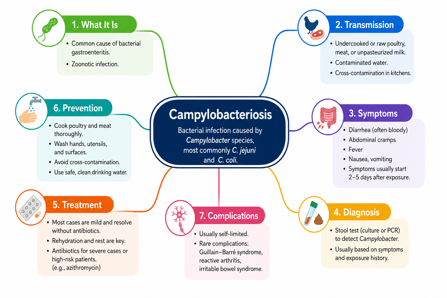
Campylobacteriosis is a bacterial gut infection — usually acquired from undercooked poultry, contaminated water, or raw milk — that causes diarrhoea, stomach cramps, and fever. Most people recover without antibiotics within a week. With an estimated 96 million cases worldwide annually, it is one of the most common foodborne illnesses globally [3]. Rare but serious complications include Guillain-Barré syndrome (GBS) [9,10].
- Campylobacter jejuni is responsible for the majority of human campylobacteriosis cases globally [6,8].
- Poultry — particularly undercooked chicken — is the primary transmission source [3,6,10].
- Symptoms typically begin 2–5 days after exposure and resolve in most patients within one to two weeks.
- Stool culture remains the diagnostic gold standard; molecular (PCR) methods are increasingly used alongside it [5,8].
- Most patients need only rehydration and rest; azithromycin or erythromycin is used when antibiotics are indicated [9,10].
- Fluoroquinolone resistance is a growing public health concern in many regions [3,9,10].
- Campylobacter is the most frequent antecedent infection identified in Guillain-Barré syndrome cases [10].
- Emerging research suggests gut microbiome composition may influence infection outcomes, but these findings are preliminary [1].
What Is Campylobacteriosis?
Campylobacteriosis is an intestinal infection caused by bacteria of the genus Campylobacter. The predominant culprit in human disease is Campylobacter jejuni, described as one of the most frequently diagnosed bacterial causes of diarrhoeal illness in the United States and worldwide [8,10]. Campylobacter coli is the second most commonly implicated species in human gastroenteritis [3,6]. Other species — including C. concisus and C. ureolyticus — are gaining recognition as clinically relevant, though their contribution to overall disease burden is smaller [6].
Globally, campylobacteriosis is estimated to cause approximately 96 million cases per year [3]. Incidence and prevalence have increased in both developed and developing countries over the past decade [6]. In North America, Europe, and Australia the rise has been particularly notable; in parts of Africa, Asia, and the Middle East the disease is endemic, especially among young children [6].
How Does It Occur? (Pathophysiology)
After ingestion, Campylobacter survives passage through the stomach and colonises the small and large intestines. Using flagella, the bacteria penetrate the intestinal mucus layer, adhere to epithelial cells, and invade them — triggering an inflammatory response that disrupts the gut lining. This leads to fluid secretion into the bowel (diarrhoea), immune activation (fever), and intestinal cramping. Bacterial toxins contribute directly to epithelial cell damage [8].
In a small proportion of cases — more likely in immunocompromised individuals — bacteria may breach the gut lining and enter the bloodstream, causing bacteraemia and potentially extraintestinal infection [8,9].
At the microbiome level, an individual’s existing gut bacterial community can influence how C. jejuni infection progresses. Recent research identified a subgroup of Sutterella wadsworthensis that produces an IgA protease capable of digesting human IgA1 and IgA2 antibodies. In laboratory models, this enzyme altered the trajectory of C. jejuni infection in human epithelial cells and modified neutrophil phagocytosis, suggesting that host microbiome composition may shape infection outcomes [1]. These findings are preliminary and have not yet been translated into clinical practice.
Causes and Transmission
Campylobacter colonises the intestines of many animals — particularly birds — without causing illness in the host. Human infection occurs when bacteria are transferred to people via several well-documented routes.
-
Contaminated poultry (primary route)
Poultry is the principal reservoir and transmission source of Campylobacter to humans [3,6]. Bacteria residing harmlessly in chicken intestines contaminate meat during slaughter and processing. Eating undercooked chicken or cross-contaminating other foods with raw poultry juices are the leading risk factors for human infection [10]. -
Eggs
The contribution of eggs to campylobacteriosis is still being characterised. A German study found Campylobacter DNA on approximately 80% of pooled eggshell samples by PCR, but viable bacteria were detected in only 11% of a subset by viability PCR and in just 6.6% by culture. The authors note that most bacteria were likely dead due to dryness, and that transfer during shell opening appeared to be limited — though infectious potential of viable-but-non-culturable bacteria requires further study [5]. -
Contaminated water
Drinking untreated or inadequately treated water — from rivers, private wells, or overwhelmed municipal systems — can transmit Campylobacter [6]. -
Unpasteurised milk and dairy products
Raw milk is a recognised vehicle for transmission; faecal contamination during milking can introduce bacteria [6,7]. -
Contact with animals
Pets (especially puppies and kittens with diarrhoea), farm animals, and urban wildlife can carry and shed Campylobacter. A study of two urban gull species in Barcelona detected Campylobacter spp. in Audouin’s gulls — whose foraging ecology exposed them to marine and coastal environments — illustrating how wildlife movement can distribute the pathogen across landscapes [4]. -
Person-to-person transmission
Direct spread is less common but can occur, particularly in households with young children or care settings where hygiene lapses arise [6]. -
International travel
Travel to regions where campylobacteriosis is endemic substantially increases risk [6].
Risk Factors
- Eating undercooked poultry or handling raw chicken without adequate hand hygiene [10]
- Drinking untreated or unfiltered water [6]
- International travel, particularly to endemic regions [6]
- Young children (under 5 years), whose developing immune systems increase susceptibility [6]
- Older adults, in whom age-related immune decline raises risk of severe disease
- Immunocompromised individuals — including those with HIV, haematological malignancies, or on immunosuppressant therapy — who face greater risk of bacteraemia and extraintestinal infection [8,9]
- Contact with farm animals or pets with diarrhoea [7]
- Consuming unpasteurised milk or dairy products [6]
- Occupational exposure in food processing, farming, or veterinary work
Symptoms
Symptoms typically appear 2–5 days after exposure and include the following:
-
Diarrhoea (often bloody)
The hallmark symptom. Inflammation and disruption of the intestinal lining cause fluid secretion into the gut. In severe cases, mucosal injury leads to blood in the stool [8]. -
Abdominal cramps and pain
Inflammatory spasm of the intestinal musculature produces colicky pain that can occasionally be severe enough to mimic appendicitis [8]. -
Fever
A systemic immune response to bacterial invasion; fever is a common feature of the acute illness [7]. -
Nausea and vomiting
Present in some patients, particularly early in the illness, due to bacterial toxins and generalised gut disruption. -
Headache and muscle aches
Flu-like systemic symptoms driven by circulating inflammatory mediators [7]. -
Dehydration
A direct consequence of ongoing diarrhoea and vomiting; most dangerous in young children, elderly people, and those unable to maintain oral intake.
Most cases resolve within 5–10 days without specific treatment. Rarely, serious complications develop:
- Guillain-Barré syndrome (GBS): A post-infectious neurological condition in which the immune system attacks peripheral nerves. Campylobacter infection is the most frequent antecedent infection identified in GBS [10].
- Reactive arthritis: Joint inflammation that can follow gastrointestinal infection [9].
- Bacteraemia and extraintestinal infection: Uncommon in immunocompetent individuals but more likely in those with immune deficiency [8,9].
- Bloody diarrhoea that persists beyond 24–48 hours
- High fever that does not improve
- Signs of dehydration: dry mouth, markedly reduced urination, extreme dizziness
- Diarrhoea lasting more than 7 days
- Weakness, numbness, or tingling in the limbs (potential early signs of GBS)
- Symptoms in a very young child, elderly person, or immunocompromised individual
- Inability to keep any fluids down
Differential Diagnosis
The clinical presentation of campylobacteriosis overlaps substantially with several other conditions. Laboratory confirmation is required to distinguish between them.
-
Salmonellosis
Caused by Salmonella species; presents with fever, diarrhoea, and cramps, also strongly linked to poultry and contaminated food. Stool culture or PCR differentiates the two [7]. -
Shigellosis (bacillary dysentery)
Shigella causes bloody diarrhoea with mucus, fever, and severe cramping closely mirroring severe campylobacteriosis. It is highly contagious and typically more explosive in onset. -
Escherichia coli gastroenteritis
Enterotoxigenic strains cause watery diarrhoea; enterohaemorrhagic strains (e.g., O157:H7) cause bloody diarrhoea and can progress to haemolytic uraemic syndrome — making identification clinically important. -
Viral gastroenteritis (norovirus, rotavirus)
Watery diarrhoea, nausea, vomiting, and sometimes fever. Tends to resolve faster (24–72 hours) and rarely causes blood in stool — useful distinguishing features. -
Inflammatory bowel disease (IBD)
Bloody diarrhoea and cramping can mimic — or be masked by — an IBD flare. Campylobacter infection may also trigger the first presentation of IBD. Colonoscopy may be needed in ambiguous cases. -
Appendicitis
Severe right lower quadrant pain in campylobacteriosis can convincingly mimic appendicitis; documented cases of surgical exploration for presumed appendicitis later attributed to Campylobacter exist [8]. -
Yersiniosis
Yersinia enterocolitica causes fever, diarrhoea (sometimes bloody), and right lower quadrant pain that can mimic both campylobacteriosis and appendicitis; also a food- and animal-borne pathogen [7].
Diagnosis
Stool culture (gold standard)
A fresh stool sample is plated on selective culture media and incubated at 42°C in a microaerophilic atmosphere — conditions that favour Campylobacter growth while suppressing other organisms. Colonies are identified biochemically and, increasingly, by MALDI-TOF mass spectrometry, which provides rapid, highly accurate species-level identification [2,8]. Antibiotic susceptibility testing performed on cultured isolates is clinically important given regional variation in resistance patterns [3,9].
Molecular testing (PCR)
PCR-based assays detect Campylobacter DNA directly from stool with high sensitivity and speed, often faster than culture. Multiplex panels simultaneously screen for multiple gut pathogens. Viability PCR (vPCR) can distinguish live from dead bacteria — relevant when assessing true infectious risk from sources such as eggshells [5]. A limitation of standard PCR is that it detects DNA from dead organisms as well as live ones, which can complicate clinical interpretation.
Supporting investigations
- Full blood count: Leukocytosis may indicate bacterial infection and immune activation.
- C-reactive protein / ESR: Elevated in bacterial gastroenteritis, though non-specific.
- Blood culture: Reserved for severely ill or immunocompromised patients in whom bacteraemia is suspected [8,9].
- Colonoscopy / sigmoidoscopy: Not routinely indicated; considered when IBD must be excluded or diagnosis remains uncertain.
Treatment
Supportive care (for most patients)
The majority of immunocompetent patients recover without antibiotics. Supportive management is the cornerstone of care:
- Oral rehydration: Replacing fluids and electrolytes is essential. Oral rehydration solutions (ORS) containing the appropriate balance of salts and glucose are preferred over plain water alone.
- Intravenous fluids: Required when dehydration is severe or oral intake cannot be maintained.
- Rest.
- Antipyretics: Paracetamol (acetaminophen) for fever and body aches. NSAIDs should be used cautiously given gastrointestinal irritation risk in the context of active diarrhoea.
- Antidiarrhoeals (e.g., loperamide): Generally avoided in bloody diarrhoea, where slowing intestinal motility may be harmful.
Antibiotic treatment
Antibiotics are considered when illness is severe or prolonged, when the patient is immunocompromised, or when bacteraemia is present or suspected [9,10]. Indications include:
- Severe or prolonged illness (symptoms lasting more than 7 days)
- Bloody diarrhoea with high fever
- Immunocompromised patients
- Bacteraemia or extraintestinal infection
- Elderly patients or very young children with significant illness
Azithromycin (a macrolide) is the preferred agent in many guidelines, inhibiting bacterial protein synthesis and generally well-tolerated [9]. Erythromycin, another macrolide, is an established alternative — particularly used in children and during pregnancy [9,10]. Specific dosing should be determined by the treating clinician based on patient weight, age, and local guidelines.
Antimicrobial resistance — a growing concern
Campylobacter resistance to multiple antibiotic classes is a serious and escalating public health problem [3,9,10]. Fluoroquinolones (e.g., ciprofloxacin) were formerly a common treatment choice, but high resistance rates have developed in many parts of the world — a trend closely linked to fluoroquinolone use in veterinary and agricultural settings [10]. Prescribing decisions should always be guided by local antibiogram data and clinical severity. Options in resistant or severe cases include:
- Fluoroquinolones (e.g., ciprofloxacin): Used in some settings, but local resistance rates must be checked before prescribing [3,10].
- Tetracyclines (e.g., doxycycline): An alternative where susceptibility is confirmed; resistance is also emerging in this class [9].
- Carbapenems: Reserved for severe, multidrug-resistant cases in hospitalised patients.
Phage biocontrol (experimental)
Bacteriophages — viruses that specifically kill bacteria — are under active investigation as an alternative to antibiotics for controlling Campylobacter. Campylobacter group II phages have shown promise in reducing bacterial loads in poultry at both preharvest and postharvest stages in controlled studies. However, consistent performance under commercial conditions requires further large-scale validation, and their clinical application in human patients is not yet established [3].
Prevention
- Cook poultry thoroughly to safe internal temperatures and avoid cross-contamination with raw meat juices.
- Wash hands thoroughly after handling raw poultry, after contact with animals, and before food preparation.
- Drink only treated or filtered water; use bottled water in settings where water quality is uncertain.
- Avoid unpasteurised milk and dairy products.
- Exercise caution when travelling to endemic regions; attend to food and water safety.
- Practise careful hand hygiene when in contact with pets or farm animals, especially those with diarrhoea.
Evidence in Context
Most clinical guidance on campylobacteriosis rests on well-established evidence, but several important gaps and uncertainties deserve acknowledgement:
- Diagnostic sensitivity: Stool culture, while the gold standard, has imperfect sensitivity and requires specific laboratory conditions (selective media, 42°C incubation, microaerophilic atmosphere). PCR is more sensitive but detects DNA from non-viable organisms. Viability PCR helps bridge this gap but is not yet universally available [5,8].
- Egg transmission: The role of eggs as an infectious source remains incompletely characterised. The largest study to date found Campylobacter DNA on approximately 80% of eggshells, but viable bacteria were confirmed in far fewer samples and culturability declined with egg age. The infectious potential of viable-but-non-culturable bacteria is uncertain and needs further investigation before definitive risk conclusions can be drawn [5].
- Microbiome interactions: The discovery that Sutterella wadsworthensis IgA protease activity can alter C. jejuni infection in cell and neutrophil models is intriguing, but these are in-vitro and primary-cell findings [1]. Their relevance to infection outcomes in living humans is unknown and not applicable to clinical practice at this time.
- Wildlife as reservoirs: Studies such as the Barcelona gull investigation [4] add nuance to understanding environmental transmission, but the relative contribution of wildlife to human case burden compared with poultry and food chain routes has not been formally quantified.
- Antimicrobial resistance surveillance: Resistance data vary significantly by country, species, and clinical setting. Inconsistent global surveillance makes universally applicable treatment recommendations difficult; local antibiogram data are essential [3,9,10].
- Phage therapy: Group II phages show genuine promise for reducing Campylobacter contamination in the food chain. However, commercial-scale validation, regulatory frameworks, and human clinical trial data are still lacking [3].
- Veterinary species: Campylobacter bilis, previously documented only in Australia and the USA as a cause of spotty liver disease in laying hens, was recently identified for the first time in the Netherlands [2]. This finding is relevant to veterinary and food-safety surveillance, but its human health implications are not yet established.
- Do I actually need antibiotics, or is this likely to resolve on its own?
- Should a stool sample be sent for culture and antibiotic sensitivity testing?
- Given local resistance patterns, which antibiotic would be most appropriate if treatment is needed?
- Am I at higher risk for complications such as Guillain-Barré syndrome?
- What warning signs should prompt me to seek care urgently?
- How can I prevent spreading this infection to household members?
- When is it safe to return to work, school, or food handling?
References
- Majzoub ME, Santiago FS, Raich SS, et al. Immunoglobulin A protease from Sutterella wadsworthensis modifies outcome of infection with Campylobacter jejuni and is associated with microbiome diversity. Gut Microbes. 2026;18(1):2611543. doi:10.1080/19490976.2025.2611543. PMID: 41496502.
- van Engelen E, Koene M, Molenaar RJ, Heuvelink A. Campylobacter bilis caused spotty liver syndrome in laying hens in the Netherlands. Vet Q. 2026;46(1):2628724. doi:10.1080/01652176.2026.2628724. PMID: 41689423.
- Rafiq MS, Haider SS, Li Z, et al. The role of Campylobacter group II phages in mitigating Campylobacter contamination across the poultry food chain: current applications and future prospects. Food Res Int. 2026;237:119365. doi:10.1016/j.foodres.2026.119365. PMID: 42169327.
- Martín-Vélez V, Yagüe de Santos S, Montalvo T, et al. Zoonotic bacterial dynamics in two sympatric urban gull species within anthropized environments. Environ Pollut. 2026;402:128316. doi:10.1016/j.envpol.2026.128316. PMID: 42142728.
- Goenaga JC, Buhler C, Wenning M, et al. Detection and quantification of thermotolerant Campylobacter spp. on eggshells by cultivation and viability real-time PCR. Int J Food Microbiol. 2026;456:111807. doi:10.1016/j.ijfoodmicro.2026.111807. PMID: 42019234.
- Kaakoush NO, Castaño-Rodríguez N, Mitchell HM, Man SM. Global epidemiology of Campylobacter infection. Clin Microbiol Rev. 2015;28(3):687–720. doi:10.1128/CMR.00006-15. PMID: 26062576.
- Chlebicz A, Śliżewska K. Campylobacteriosis, salmonellosis, yersiniosis, and listeriosis as zoonotic foodborne diseases: a review. Int J Environ Res Public Health. 2018;15(5):863. doi:10.3390/ijerph15050863. PMID: 29701663.
- Fitzgerald C. Campylobacter. Clin Lab Med. 2015;35(2):289–298. doi:10.1016/j.cll.2015.03.001. PMID: 26004643.
- Moore JE, Corcoran D, Dooley JS, et al. Campylobacter. Vet Res. 2005;36(3):351–382. doi:10.1051/vetres:2005012. PMID: 15845230.
- Fields PI, Swerdlow DL. Campylobacter jejuni. Clin Lab Med. 1999;19(3):489–504. PMID: 10549422.