Clubfoot (congenital talipes equinovarus) is a common birth condition in which the foot is twisted inward and downward. It affects approximately 1–2 per 1,000 live births worldwide. [9][10] The Ponseti method — serial casting followed by bracing — is the gold standard treatment, and children who complete the full protocol, including the bracing phase, can achieve good long-term function. [6][7]
- Clubfoot is one of the most common congenital musculoskeletal conditions globally, affecting approximately 1–2 per 1,000 live births. [9][10]
- It can often be detected before birth on second-trimester ultrasound. [9]
- The Ponseti method — serial casting, Achilles tenotomy in most cases, and prolonged bracing — is the accepted gold standard. [6][7][10]
- An accelerated casting schedule (more frequent cast changes) achieves comparable correction rates with shorter total treatment duration, though long-term follow-up data are still limited. [1]
- Brace-related complications (skin redness, blisters, swelling) are common in the early bracing phase and require proactive caregiver education and follow-up. [2]
- Family brace compliance is a critical determinant of long-term success; premature discontinuation is strongly associated with relapse. [7]
- Children with idiopathic clubfoot treated with Ponseti can achieve quality-of-life scores comparable to healthy peers in older age groups, though caregiver burden peaks in the early bracing years. [3]
What Is Clubfoot?
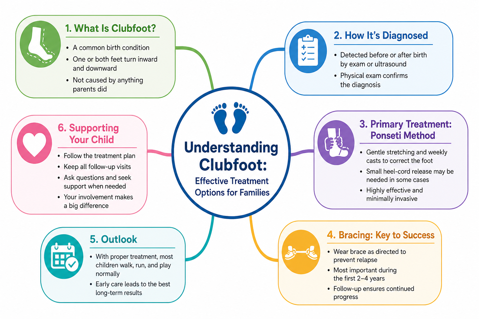
Clubfoot — medically termed congenital talipes equinovarus (CTEV) — is a structural foot deformity present from birth in which the foot is turned inward and downward and cannot be passively corrected to a neutral position. The deformity has four recognisable components: cavus (high midfoot arch), adductus (forefoot turning inward), varus (heel turned inward), and equinus (heel pulled upward). [9]
It is one of the most frequently encountered musculoskeletal birth conditions, affecting approximately 1 to 2 babies per 1,000 live births worldwide. [9] In the United Kingdom, approximately 1 in 1,000 births is affected. [10] Boys are affected more often than girls, and roughly half of cases involve both feet. When left untreated, clubfoot persists into adult life and is associated with significant functional limitation and reduced quality of life. [7]
How Does Clubfoot Develop?
The precise mechanism is incompletely understood. During fetal development, the talus bone is abnormally positioned — rotated inward and downward — and the tendons on the posteromedial aspect of the foot, particularly the Achilles tendon and posterior tibialis, become shortened and contracted. Ligaments and joint capsules on the inner and posterior foot tighten, and in more severe cases bony morphology is also altered. [6][9]
The four structural components — cavus, adductus, varus, and equinus — together produce the characteristic appearance identifiable on prenatal ultrasound and at birth. [9]
Causes and Risk Factors
Clubfoot is considered multifactorial in origin. In the majority of cases no identifiable cause is found (idiopathic clubfoot), and these respond best to conservative treatment. [6][7] Recognised associations include:
- Idiopathic: The most common form. The baby is otherwise healthy and no underlying condition is identified. [6][7]
- Neuromuscular conditions: Spina bifida, arthrogryposis, and other disorders affecting muscle and nerve development are strongly associated with clubfoot.
- Chromosomal and syndromic conditions: Clubfoot has been reported in association with trisomy 18 [8] and Turner mosaicism. [4] These syndromic cases may present distinct management challenges.
- Restricted fetal movement: Oligohydramnios (reduced amniotic fluid) limits fetal movement and may mechanically contribute to abnormal foot positioning.
- Family history: A first-degree family history of clubfoot increases risk.
- Male sex: Boys are affected approximately twice as often as girls.
- Maternal smoking: Some evidence links smoking in pregnancy — particularly in families with genetic predisposition — to higher risk.
It is important to note that specific risk percentages beyond those stated in cited sources are not reproduced here, as population estimates vary considerably across studies.
Symptoms and Clinical Features
Clubfoot is usually apparent immediately at birth. Key features include:
- Foot rotated inward and downward: The sole faces inward or upward, reflecting the malpositioned talus and contracted posteromedial tendons. [9]
- High arch (cavus): Exaggerated midfoot arch from abnormal tension in the plantar structures.
- Smaller foot and calf: The affected limb is often slightly smaller due to reduced intrauterine muscle activity.
- Rigidity: Unlike postural deformities, true clubfoot resists passive correction. This distinguishes it clinically from flexible positional variants.
- Achilles tendon tightness: The heel cannot be brought to a plantigrade position because the Achilles tendon is shortened.
- Medial skin creases: Excess skin folds on the inner border of the foot reflect the compressed tissue configuration.
Clubfoot itself does not typically cause pain in infancy. However, an uncorrected clubfoot walking on a deformed surface causes pain, pressure sores, and significant functional limitation in later childhood and adult life. [7]
Differential Diagnosis
Several conditions can resemble clubfoot. Distinguishing them is clinically important because management differs:
- Metatarsus adductus: The forefoot turns inward but the heel is normally positioned. Usually flexible and often resolves spontaneously.
- Positional (postural) clubfoot: The foot appears clubbed but corrects fully with gentle manual pressure. No structural deformity is present.
- Congenital vertical talus (rocker-bottom foot): The talus is vertically oriented — the opposite direction from clubfoot. It is rare, rigid, and requires different treatment.
- Calcaneovalgus foot: The foot points outward and upward. Usually positional and self-resolving.
- Arthrogryposis-associated deformity: Foot deformity is part of a broader syndrome of joint contractures and requires a tailored approach.
Diagnosis
Diagnosis is primarily clinical. Severity is graded using structured scoring tools such as the Pirani score (0–6 scale) or the Diméglio classification, which guide cast number estimates and treatment planning. [1][2]
Prenatal Ultrasound
Clubfoot is frequently detected on routine second-trimester ultrasound. A fetal foot seen in a fixed abnormal position relative to the lower leg raises suspicion. [9] Prenatal detection enables early specialist counselling and planning for prompt postnatal treatment.
Plain Radiography
X-rays reveal the positional relationships of the tarsal bones — particularly the talus and calcaneus — which are characteristically malaligned in clubfoot. [9] Specific angular measurements on anteroposterior and lateral views help confirm the diagnosis and monitor correction. Because much of the infantile foot skeleton remains cartilaginous, radiographs become more informative after some ossification has occurred.
Ultrasound
Ultrasound visualises cartilaginous structures that are invisible on X-ray, carries no radiation exposure, and is well suited for monitoring the response to Ponseti casting in young infants. [9]
MRI
MRI provides the most comprehensive view of bone, cartilage, tendons, and ligaments together. [9] It is not required for routine idiopathic clubfoot but is valuable for complex, recurrent, or syndromic cases where detailed surgical planning is needed.
Treatment
Treatment should begin as early as possible. Neonatal tissues are highly malleable, and early intervention takes advantage of this biological window. The overarching goal is a plantigrade, pain-free, functional foot. [6][7]
The Ponseti Method — Gold Standard
The Ponseti method is the most widely accepted treatment for idiopathic congenital clubfoot worldwide. [6][7][10] It consists of three phases:
- Serial casting: Beginning ideally within the first weeks of life, the clinician gently manipulates the foot toward a corrected position and immobilises it with a plaster or fiberglass cast from toes to thigh. Casts are changed at weekly intervals (or more frequently in the accelerated protocol — see below) as the foot progressively corrects. Most infants require approximately 5–7 casts. [1][7]
- Percutaneous Achilles tenotomy: In the majority of cases the Achilles tendon is too contracted to allow full correction by casting alone. A minor percutaneous tenotomy is performed under local anaesthetic to release it, after which a final cast is worn for approximately three weeks while the tendon heals. [6][7]
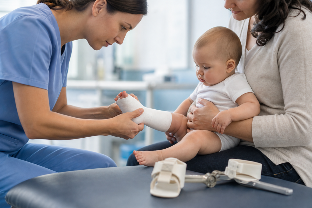
- Foot abduction orthosis (FAO) bracing: After casting, the child wears a foot abduction brace — a bar connecting two shoes that hold the feet in an outwardly rotated position. The brace is worn for extended periods (initially full-time, then reducing to night-time and nap-time use) for several years. [2][5] This phase is critical: premature discontinuation is the primary driver of relapse. [7]
Accelerated Ponseti Protocol
A systematic review and meta-analysis of 16 studies involving 957 patients found that an accelerated casting schedule (cast changes every 5 days rather than weekly) produced outcomes statistically equivalent to the standard protocol in post-treatment Pirani score, tenotomy rate, and relapse rate. The accelerated group required slightly more total casts but completed casting in a significantly shorter total duration (mean approximately 20 days shorter). [1] The authors noted that blinding was generally lacking across studies and that standardised implementation and long-term follow-up are recommended before this protocol is considered routine. [1]
Brace-Related Complications
The bracing phase is associated with a notable burden of complications. In a prospective Pakistani series of 91 infants, skin reddening (75.8%) and pain (88%) were the most frequently reported problems in the first 24 hours of bracing, followed by blisters or sores (42.9%) and distal tibial swelling (26.4%) by day 7. Muscle atrophy or weakness peaked at 32% by day 14 and persisted in 24.2% of patients at 30 days. Despite these complications, brace compliance was high and no relapses occurred during the 6-month follow-up. [2] Importantly, the authors note a scarcity of focused research in this area and call for improved brace design, personalised follow-up, and effective caregiver guidance — particularly in low-resource settings. [2]
A separate study examining the uninvolved leg in children with unilateral clubfoot found statistically significant differences in external hip rotation and external bimalleolar axis compared to normative controls, but no significant impact on thigh-foot angle, foot progression angle, or pedobarographic foot loading. The authors concluded these differences are clinically insignificant and that families can be reassured the brace does not cause meaningful deformity in the uninvolved limb. [5]
Quality of Life and Caregiver Burden
A cross-sectional survey of 180 children treated with the Ponseti method in China found that children aged 0–1 year showed reduced physical and social functioning scores compared to healthy references, whereas children aged 5–18 years scored significantly higher than healthy references on most quality-of-life domains. Caregiver burden (measured by the Zarit Burden Interview) was highest during active treatment and declined after age five years; 54.4% of caregivers reported no or little burden overall. Factors associated with better patient quality of life included older age and fewer treatment-related complications; caregiver quality of life was associated with brace adherence. [3] These findings support the importance of complication prevention and ongoing brace adherence education during clinic visits. [3]
When the Ponseti Method Is Insufficient — Additional Interventions
For relapsed, neglected, or syndromic clubfoot, additional interventions may be required:
- Repeat casting: Mild early relapses can often be re-corrected with a further series of Ponseti casts.
- Anterior tibialis tendon transfer (ATTT): For children (typically over 2–2.5 years) with dynamic forefoot supination due to muscle imbalance, surgical relocation of the anterior tibialis tendon to a more central position on the foot can rebalance the deforming force. [6]
- Posteromedial release (PMR) surgery: Extensive soft-tissue release was the dominant treatment before Ponseti became established. It is now reserved for severe, rigid, or multiply recurrent deformities unresponsive to conservative management, and carries higher long-term risks of stiffness and arthritis. [6]
- Ilizarov external fixation: Used in neglected or previously operated severe clubfoot, where gradual mechanical distraction repositions the foot over weeks to months.
Evidence in Context: What We Know and What Remains Uncertain
The evidence base for the Ponseti method is substantial and consistently favourable for idiopathic clubfoot, supported by clinical reviews, a Pediatrics clinical report, UK consensus guidelines, and a growing body of prospective data. [6][7][10] Key limitations and uncertainties to keep in mind:
- Idiopathic vs. syndromic clubfoot: Most evidence, including the meta-analysis of the accelerated protocol, pertains to idiopathic cases. Evidence for syndromic or neurogenic clubfoot is less robust and less generalisable. [1][6]
- Long-term outcomes: Follow-up data extending beyond 10 years remain limited. The accelerated Ponseti protocol specifically lacks long-term outcome data, and the meta-analysis authors recommend standardised implementation before wide adoption. [1]
- Brace complications: These are common but under-studied. The available prospective data come from a single clinic in a low-resource setting and may not generalise to all populations. Further research on brace design and complication management is needed. [2]
- Caregiver burden data: The quality-of-life and caregiver burden study was cross-sectional, conducted by online survey in WeChat groups, and limited to one country; selection bias is possible. [3]
- Uninvolved limb effects: The observed rotational differences in the uninvolved limb are statistically but not clinically significant based on current data from a single retrospective cohort. [5]
- If clubfoot is suspected on prenatal ultrasound, seek referral to a pediatric orthopedic specialist before birth so treatment can begin immediately after delivery.
- If your newborn’s foot appears twisted inward or downward and cannot be gently moved to a neutral position.
- If brace-related skin redness does not resolve within 20–30 minutes of brace removal, or if blisters, open sores, or persistent swelling develop. [2]
- If your child develops a limp, seems in pain, or the foot appears to be drifting back toward its original position — possible signs of relapse. [7]
- If your child is having difficulty tolerating the brace and compliance is at risk; early intervention from the treatment team may prevent relapse.
- Is my child’s clubfoot idiopathic, or should we investigate an underlying chromosomal or neuromuscular condition?
- How many casts will my baby likely need, and would an accelerated casting schedule (more frequent changes) be appropriate in our situation?
- What brace complications should I watch for, and at what point should I contact the clinic?
- What are the early signs of relapse, and what should I do if I notice them?
- At what point might surgery be considered, and what would that involve?
- How long does my child need to wear the brace, and what happens if we miss nights of wear?
- Are there support organisations or resources for families managing clubfoot treatment?
References
- Llombart-Blanco R, Mariscal G, Altabbaa H, Polevoi E, Barrios C, Llombart-Ais R. Accelerated versus standard Ponseti method for idiopathic clubfoot: A systematic review and meta-analysis of efficacy and safety. Foot Ankle Surg. 2026 Jul;32(5):433–441. doi: 10.1016/j.fas.2025.12.010. PMID: 41549019.
- Khattak HK, Qazi AJ, Khan EW, Ahmad E, Khan ZA, Noormal, Abubakkar S, Hayat S. Complications Arising From the Use of Foot Abduction Orthosis (FAO) in Ponseti-Treated Clubfoot Patients. J Pediatr Orthop. 2026 Jul 1;46(6):e564–e570. doi: 10.1097/BPO.0000000000003153. PMID: 41272987.
- Yang Y, Yang X, Guan L, Xu J, Chen W, Bai G. Health-related quality of life and caregiving burden in pediatric clubfoot patients treated with Ponseti method in China: a cross-sectional study. BMC Musculoskelet Disord. 2026 Jun 12. doi: 10.1186/s12891-026-10075-w. PMID: 42286573.
- Luo X, Niu H, Tang Y, Xu J, Zhou F, Cong X, et al. Clinical analysis of prenatal diagnosis, ultrasound findings, and follow-up in 92 Turner mosaic fetuses: a retrospective analysis of 12-year experience. BMC Pregnancy Childbirth. 2026 Jun 12. doi: 10.1186/s12884-026-09453-y. PMID: 42286522.
- Lawing CR, Bailey CE, Garg S, Aguilera Flores J, Bailey JM, Saraswat P, Westberry DE. Evaluation of rotational profile and foot loading in the uninvolved leg of children with unilateral clubfoot treated with foot abduction orthoses. J Pediatr Orthop B. 2026 Jun 3. doi: 10.1097/BPB.0000000000001357. PMID: 42228848.
- Rieger MA, Dobbs MB. Clubfoot. Clin Podiatr Med Surg. 2022 Jan;39(1):1–14. doi: 10.1016/j.cpm.2021.08.006. PMID: 34809788.
- Cady R, Hennessey TA, Schwend RM. Diagnosis and Treatment of Idiopathic Congenital Clubfoot. Pediatrics. 2022 Feb 1;149(2):e2021055555. doi: 10.1542/peds.2021-055555. PMID: 35104362.
- Pachajoa H. Double aneuploidy (trisomy X, trisomy 18) in a newborn with trisomy 18 phenotype [Article in Spanish]. Arch Argent Pediatr. 2013 Jul–Aug;111(4):e101–4. doi: 10.5546/aap.2013.e101. PMID: 23912296.
- do Amaral E Castro A, Peixoto JB, Miyahara LK, Akuri MC, Moriwaki TL, Sato VN, et al. Clubfoot: Congenital Talipes Equinovarus. Radiographics. 2024 Jul;44(7):e230178. doi: 10.1148/rg.230178. PMID: 38935547.
- Hopwood S, Khan F, Kemp J, Rehm A, Ashby E. Clubfoot: an overview and the latest UK guidelines. Br J Hosp Med (Lond). 2023 Jan 2;84(1):1–7. doi: 10.12968/hmed.2022.0380. PMID: 36708340.