Cleft lip is a congenital facial difference in which the upper lip does not fully fuse during early pregnancy. It is one of the most common craniofacial birth defects worldwide and is managed primarily through surgery, with multidisciplinary support continuing from infancy into adulthood.
Key Takeaways
- Cleft lip results from failure of lip tissues to fuse during the first trimester, typically between weeks 4 and 7 of gestation.
- A 2022 systematic review and meta-analysis found a global prevalence of cleft lip of 0.3 per 1,000 live births, and cleft lip combined with cleft palate of 0.45 per 1,000 live births [6].
- Both genetic and environmental factors contribute; most cases are non-syndromic and multifactorial [7][8].
- Surgical repair (cheiloplasty) is the primary treatment; timing and technique vary by centre [9][10].
- Early pre-surgical interventions such as nasoalveolar molding (NAM), commenced within one month of birth, may improve surgical outcomes [9].
- A multidisciplinary team approach is essential from birth through adulthood [8][10].
- Psychosocial outcomes after treatment are variable and not universally positive; ongoing psychological support is important [1].
What Is Cleft Lip?
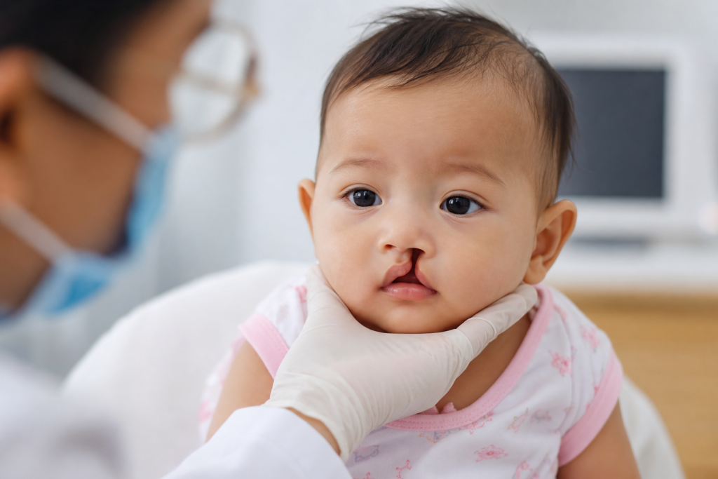
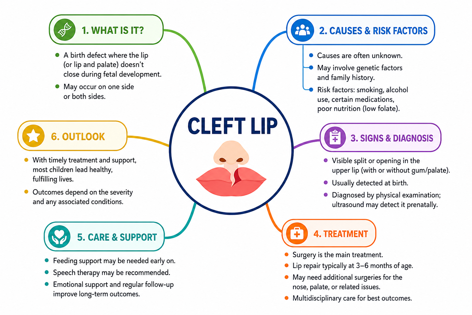
Cleft lip is a gap or split in the upper lip present from birth, arising when the lip tissues fail to fuse during fetal development. The opening ranges from a small notch to a complete gap extending into the nose. It may affect one side (unilateral) or both sides (bilateral), and frequently co-occurs with cleft palate, though it can occur in isolation.
A 2022 systematic review and meta-analysis of studies covering more than 17 million individuals reported a global prevalence of cleft lip of 0.3 per 1,000 live births (95% CI: 0.26–0.34), and cleft lip with palate of 0.45 per 1,000 live births (95% CI: 0.38–0.52) [6]. A 2009 Lancet seminar reported that all oral cleft forms combined arise in approximately 1.7 per 1,000 liveborn babies, with notable ethnic and geographic variation [7]. The condition appears slightly more frequently in males and in Asian and Indigenous populations [7].
How Does It Occur?
The face is assembled from separate tissue segments that must grow toward each other and fuse. The upper lip forms through the merging of the medial nasal processes and the maxillary processes, a process that occurs between approximately weeks 4 and 7 of gestation. When this fusion is disrupted — by genetic, nutritional, or environmental factors — a cleft results [7].
At a cellular level, disruptions in neural crest cell migration, programmed cell death (apoptosis), and epithelial-mesenchymal interactions have been implicated [7][8]. Cleft lip may be isolated (non-syndromic, the most common form) or part of a broader genetic syndrome (syndromic), each with distinct underlying mechanisms [8].
Causes and Risk Factors
Cleft lip is multifactorial — genetics, environment, and their interaction all contribute. No single cause explains all cases.
Genetic Factors
Non-syndromic cleft lip is influenced by multiple genes. Variants in genes including IRF6 have been identified as significant contributors [8]. Syndromic forms — where the cleft is part of a larger genetic condition such as Van der Woude syndrome or Pierre Robin sequence — account for a substantial minority of cases [7][8]. A family history of cleft lip or palate is considered the strongest individual risk factor [7][8].
Environmental Factors
The 2009 Lancet seminar identified several environmental risk factors with supporting evidence [7]:
- Maternal smoking during the first trimester is a well-evidenced modifiable risk.
- Alcohol use during early pregnancy has been associated with increased risk.
- Certain medications — particularly anticonvulsants (e.g., phenytoin, valproate) and corticosteroids — taken during the critical window of lip formation.
- Folate deficiency — inadequate folic acid intake before and during early pregnancy is a recognised risk factor, though the magnitude of risk reduction from supplementation varies across studies [7].
- Maternal infections (e.g., rubella) and poorly controlled maternal diabetes have also been associated with increased risk [7].
Assisted Reproductive Technology (ART)
A 2026 retrospective study of 745,671 fetuses in Hunan Province, China, found that ART-conceived pregnancies had a significantly higher risk of birth defects overall (adjusted OR 1.84, 95% CI: 1.68–2.01), with cleft lip and palate among the specifically increased subtypes. Twin pregnancies mediated approximately 14.33% of the ART–birth defect association [2]. These findings require replication in other populations before firm conclusions can be drawn.
Other Risk Factors
- Ethnicity — higher rates reported in Asian and Native American populations compared to European and African populations [7].
- Male sex — cleft lip with or without cleft palate is more common in males; isolated cleft palate is more common in females [7].
Symptoms
The primary sign of cleft lip is usually visible at birth — or earlier on prenatal ultrasound — but associated features extend beyond appearance.
- A visible gap or split in the upper lip — the hallmark feature, ranging from a small notch to a complete opening extending to the nose [7].
- Difficulty feeding — the gap disrupts the oral seal required for suction; specialised feeders are often needed [10].
- Nasal deformity — flattening or asymmetry of the nose on the affected side, due to altered structural support [7].
- Speech difficulties — particularly when the palate is also involved; hypernasality and articulation errors may develop as the child grows [5][10].
- Dental abnormalities — missing, extra, or malpositioned teeth, especially when the alveolus is affected [9].
- Middle ear disease and hearing difficulties — children with associated cleft palate are at increased risk of otitis media with effusion [10].
- Psychosocial challenges — self-esteem difficulties and social challenges may arise and can persist into adulthood; outcomes are variable and not universally negative [1].
Differential Diagnosis
Several conditions may resemble or co-occur with cleft lip and should be considered during assessment [7][8][10]:
- Isolated cleft palate — involves only the palate with a normal-appearing lip; often missed without careful oral examination of all newborns [7].
- Submucous cleft palate — a cleft hidden beneath intact mucosa; clues include a bifid uvula, a notch at the posterior hard palate, and hypernasality [10].
- Van der Woude syndrome — caused by IRF6 mutations; presents with cleft lip and/or palate alongside lower lip pits; the most common syndromic cause of oral clefts [8].
- Pierre Robin sequence — a triad of micrognathia, glossoptosis, and cleft palate; respiratory difficulty is prominent [8].
- Median cleft lip — a rare midline cleft, often associated with other midline anomalies; distinguished from the more common paramedian cleft lip [7].
Diagnosis
Prenatal Detection
Cleft lip can often be detected during the routine second-trimester ultrasound, typically performed between 18 and 22 weeks of gestation. Unilateral cleft lip is frequently identifiable as a gap in the upper lip; cleft palate alone is harder to detect on standard 2D ultrasound. Three-dimensional ultrasound has been used to improve visualisation of facial structures, though detection rates depend on operator experience and fetal position [7]. Early prenatal identification enables family counselling and planning of postnatal care.
Postnatal Diagnosis
After birth, diagnosis is confirmed by direct clinical examination. Clinicians assess cleft extent (unilateral vs. bilateral, complete vs. incomplete) and whether the palate is involved. As the child grows, hearing assessments, dental radiographs, and speech evaluations are incorporated into ongoing monitoring [5][10].
Advanced Imaging for Treatment Planning
Postnatally, cone-beam CT (CBCT) and finite element analysis (FEA) are increasingly used to plan surgical and orthodontic treatment. A 2026 review described how FEA models — incorporating sutures and scar tissue — can simulate orthodontic force transmission and assess biomechanical differences between unilateral and bilateral cleft lip and palate, supporting personalised preoperative planning [3].
Treatment
Treatment of cleft lip is multidisciplinary, spanning from the neonatal period through adulthood. No single intervention is sufficient; care is coordinated across surgery, speech-language therapy, orthodontics, dentistry, and psychology [8][10].
Pre-Surgical Preparation
Nasoalveolar molding (NAM) is a non-surgical approach using a custom intraoral device to gradually reshape the nose, lip, and gum tissues before surgery. A systematic review of randomised controlled trials found that commencing NAM within one month of birth offers meaningful benefit [9], though evidence on optimal protocols continues to be gathered.
The Super-Passive Alveolar Correcting Equipment (SPACE), a molar-supported passive plate, is a newer presurgical device studied in Japan. In a cohort of 83 patients with unilateral cleft lip, alveolus, and palate, SPACE reduced the mean alveolar gap to 1.87 mm before surgery and facilitated a one-stage repair (lip repair, gingivoperiosteoplasty, and palatoplasty) at approximately 6 months of age, with no oronasal fistulas observed. At age 5, malarticulation occurred in 14.6% of the SPACE one-stage group versus 42.2% in a conventional multistage comparison group, and the secondary alveolar bone grafting avoidance rate was 38.6% [4]. These results are from a single-centre study and require broader validation.
Surgical Repair (Cheiloplasty)
Surgery is the primary treatment for cleft lip. Cheiloplasty is typically performed when the infant is medically stable, commonly between 3 and 6 months of age, though timing varies by centre and protocol [10]. The goals are to close the gap, restore lip function and appearance, and improve nasal symmetry.
Commonly used techniques include:
- Millard’s rotation-advancement technique — widely used internationally, with the scar placed along the natural philtral column.
- Fisher’s anatomic subunit repair and Mohler’s technique — refined approaches with evidence from randomised controlled trials supporting their use [9].
Choice of technique depends on cleft extent, surgeon experience, and centre protocols [9][10].
Secondary and Supportive Treatments
- Alveolar bone grafting — typically performed around ages 8–10 to fill bone gaps before permanent tooth eruption. Recombinant human bone morphogenetic protein-2 (rh-BMP2) has shown promise as an alternative to traditional autografts [9].
- Orthodontic treatment — for dental alignment and bite management, informed increasingly by biomechanical planning tools [3].
- Speech and language therapy — early, specialised assessment is important, particularly when the palate is involved. The West Midlands Assessment of Speech-Preschool (WAS-P), developed and piloted in the UK, represents an effort to standardise speech assessment at age 3 across cleft units, enabling outcome comparisons; it is currently undergoing further validation [5].
- Orthognathic surgery — for jaw misalignment persisting into adolescence or adulthood. A 2026 qualitative study of 16 adults found that many experienced increased self-confidence and social ease following orthognathic surgery, describing it as transformative; however, psychosocial empowerment was neither universal nor immediate, with some reporting neutral or context-dependent outcomes [1].
- Middle ear management — children with associated cleft palate require monitoring for otitis media with effusion and hearing difficulties [10].
- Psychological support — given the documented psychosocial challenges across the lifespan, psychological care should be integrated throughout the treatment journey [1].
Evidence in Context
While the evidence base for surgical repair of cleft lip is well established, several important caveats apply:
- Much research on specific surgical techniques comes from single-centre studies or trials with limited sample sizes. There is a recognised need for multicentre randomised controlled trials to reduce variability and identify optimal protocols [9].
- The psychosocial impact of cleft lip and its treatment is clinically significant but frequently underrepresented in quantitative outcome research. The available qualitative evidence suggests outcomes are variable and individual [1].
- Data on ART as a risk factor for cleft lip and palate derive from a single large regional dataset in China and cannot yet be generalised globally [2].
- Advanced imaging and biomechanical modelling tools (e.g., FEA) show promise for personalised treatment planning but remain areas of active development rather than established standard practice [3].
- Novel presurgical devices such as SPACE require multicentre validation before widespread adoption [4].
- Speech assessment standardisation efforts such as the WAS-P are at an early stage; the tool has not yet been fully validated [5].
Practical Guidance for Families
If your child has been diagnosed with cleft lip — prenatally or at birth — the following questions may help guide conversations with your clinical team:
- What type and extent of cleft does my child have, and is the palate involved?
- Is nasoalveolar molding appropriate, and if so, when should it begin?
- What surgical technique is planned, and what outcomes can realistically be expected?
- When should speech and language therapy assessments begin?
- What hearing monitoring will be in place?
- What psychological and family support services are available?
- What does the long-term follow-up plan involve, and who coordinates care?
Care is best provided by a coordinated multidisciplinary cleft team, which may include plastic or craniofacial surgeons, orthodontists, speech-language therapists, audiologists, dentists, geneticists, and psychologists [8][10].
References
- Lundberg J, Sjöström M, Molin J. Psychosocial empowerment following orthognathic surgery for patients born with cleft lip and/or palate. Int J Qual Stud Health Well-being. 2026;21(1):2674347. doi:10.1080/17482631.2026.2674347. PMID: 42138295.
- Hu F, Zhu H, Fang J, Li B, Xie D. Assisted reproductive technology and birth defects: analysis of the mediating pathways in twin pregnancies. J Matern Fetal Neonatal Med. 2026;39(1):2658942. doi:10.1080/14767058.2026.2658942. PMID: 42086487.
- Deng M, Wang Y. Advances in the application of finite element analysis in the diagnosis and treatment of cleft lip and palate and alveolar cleft. J Craniomaxillofac Surg. 2026;54(8):104602. doi:10.1016/j.jcms.2026.104602. PMID: 42166884.
- Oyama A, Funayama E, Okamoto T, et al. Super-Passive Alveolar Correcting Equipment (SPACE): a novel presurgical cleft device with 5-year outcomes following one-stage repair. J Craniomaxillofac Surg. 2026;54(8):104422. doi:10.1016/j.jcms.2025.12.001. PMID: 41513529.
- Fitzpatrick B, Coad J, Sell D, Price M, Rihtman T. Development and Testing of the West Midlands Assessment of Speech-Preschool (WAS-P) for 3-Year-Olds With Cleft Palate ± Cleft Lip. Int J Lang Commun Disord. 2026;61(4):e70269. doi:10.1111/1460-6984.70269. PMID: 42231836.
- Salari N, Darvishi N, Heydari M, et al. Global prevalence of cleft palate, cleft lip and cleft palate and lip: A comprehensive systematic review and meta-analysis. J Stomatol Oral Maxillofac Surg. 2022;123(2):110–120. doi:10.1016/j.jormas.2021.05.008. PMID: 34033944.
- Mossey PA, Little J, Munger RG, Dixon MJ, Shaw WC. Cleft lip and palate. Lancet. 2009;374(9703):1773–1785. doi:10.1016/S0140-6736(09)60695-4. PMID: 19747722.
- Babai A, Irving M. Orofacial Clefts: Genetics of Cleft Lip and Palate. Genes (Basel). 2023;14(8):1603. doi:10.3390/genes14081603. PMID: 37628654.
- Wadde K, Chowdhar A, Venkatakrishnan L, et al. Protocols in the management of cleft lip and palate: A systematic review. J Stomatol Oral Maxillofac Surg. 2023;124(2):101338. doi:10.1016/j.jormas.2022.11.014. PMID: 36410660.
- Worley ML, Patel KG, Kilpatrick LA. Cleft Lip and Palate. Clin Perinatol. 2018;45(4):661–678. doi:10.1016/j.clp.2018.07.006. PMID: 30396411.
“`